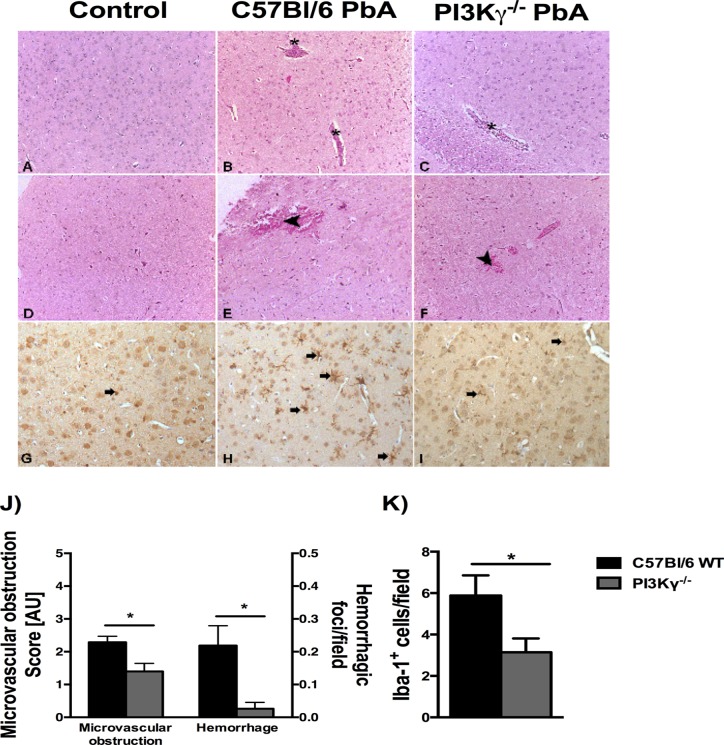
Fig 2. Infected PI3Kγ-/- mice present mild histopathological changes in brain.
Representative photomicrographs of hematoxylin/eosin (HE)-stained brain sections from uninfected control mice (A, D), or PbA-infected C57BL/6 (B, E) and PI3Kγ-/- (C, F) mice on day 6 p.i. Normal histological appearance of cerebrum (A) and brainstem (D) from uninfected control mice. Brain sections from a PbA-infected wild-type animal exhibiting intravascular leukocyte aggregates (asterisks) in cerebrum (B) and hemorrhage (arrow head) in brainstem (E). Note vascular plugging (asterisk) in the deep cerebral cortex (C) and small hemorrhagic foci (arrow head) in the brainstem (F) from PbA-infected PI3Kγ-/- animal. The severity of microvascular obstruction and cerebellar hemorrhagic areas (μm2/field) in infected mice demonstrated a significant reduction in brain alterations in PI3Kγ-/- mice (J). Immunohistochemical staining for anti-Iba-1 showing no reactive microglia (arrow) in non-infected WT mouse brain (G). Microglial cells (arrows) displaying morphological changes (thickening of the microglial processes) and immunoreactivity for Iba-1 in the cerebrum of PbA-infected wild-type animal (H) and PI3Kγ-/- mouse (I). Quantification of reactive microglia (K) confirmed lower number in brain sections of infected-PI3Kγ-/- mice when compared with PbA-infected WT animal. Significant differences were indicated by *p<0.05 (n = 5 mice per group). Original magnification: A-I: x200. Sections were captured with a digital camera (Optronics DEI-470) connected to a microscope (Olympus IX70).

