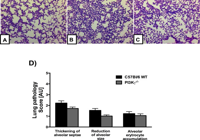
Fig 5. Lung pathology induced by PbA infection seems to be independent of PI3Kγ.
Representative photomicrographs of hematoxylin/eosin (HE)-stained lung sections from uninfected control mice (A), or PbA-infected C57BL/6 (B) and PI3Kγ-/- (C) mice on day 6 p.i. Normal histological section of lung parenchyma with standard architecture from uninfected control (A). Lung sections from a PbA-infected C57BL/6 and PI3Kγ-/- animal exhibiting slight interstitial edema, thickening of alveolar septae and a reduction of alveolar size (B, C). Semi-quantification score for histopathological changes in the lung of PbA-infected mice showed no significant difference between C57BL/6 and PI3Kγ-/- infected mice (n = 5 mice for each group) (D). Original magnification: A-D: x200. Sections were captured with a digital camera (Optronics DEI-470) connected to a microscope (Olympus IX70).

