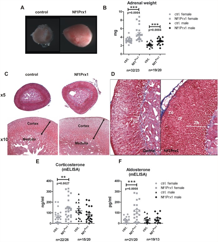
Fig 2. Increased weight of adrenal glands and defects of adrenal cortex zonation in the female Nf1Prx1 mice.
(A) Macroscopic appearance of the representative female control and mutant adrenal glands. (B) Increased adrenal gland weight in female Nf1Prx1 mice. (C) Representative H&E staining of adrenal glands from 2-month-old female wt and Nf1Prx1 mice. Higher magnification illustrating, thicker and structurally irregular and disorganized adrenal cortex in comparison with control mice (lower panel). (D) Azan stained sections. Increased thickness of the (ZR) zona reticularis and (ZF) zona fasciculata in the adrenals of mutant Nf1Prx1 mice. (E, F, G) ELISA based quantitative analysis of the serum corticosterone and Aldosterone levels. Note a female specific increase of the corticosterone and Aldosterone level (measured 9–11 a.m.) Statistical evaluation was done with T-test with Welch’s correction.

