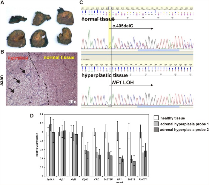
Fig 6. Loss of heterozygosity in NF1 locus associates with adrenal macronodular hyperplasia and cortisol overproduction in NF1 female patient.
A.) Surgically removed adrenal gland of the 30 years old NF1 patient with yellow coloured adrenal hyperplasia. B.) Azan stained section of the adrenal tissue showing border between hyperplastic and healthy adrenal cortex tissue. C.) Sanger sequencing showing mixed sequences of the wt allele and c.405delG mutation in the blood (same in normally part of the adrenal gland) and a loss of heterozygosity detected in the hyperplastic part of the adrenal gland. D.) Real time quantitative PCR measuring gene copy number throughout chromosme 17 and on the control chromosomes. The mean values were calculated for each amplicon relative to albumin (ALB) on 4q11 as autosomal two-copy control. Error bars indicate the 95% confidence interval as given by the ABI 7900 SDS-Software. Results were calibrated to the mean values of a healthy control tissue (white bars). Decreased signal intensities were obtained in two hyperplastic adrenal tissue samples (grey bars) for all amplicons on chromosome 17 including amplicons for NF1 type1 deletions (CPD, RHOT1), NF1 type 2 deletions (SUZ12P, SUZ12), an amplicon localized upon the NF1 mutation detected by sequencing (NF1-exon4), and an amplicon on the short arm of chromosome 17 (17p12). In contrast, control amplicons localized on chromosome 6 (6p21.1, 6q21) and the X chromosome (Xq28) revealed normal signal intensities. These results indicate that a complete loss of chromosome 17 lead to NF1 loss of heterozygosity (LOH) detected by sequencing analysis. Copy-number analyses were repeated twice with similar results and confirmed using additional qPCR amplicons (not shown).

