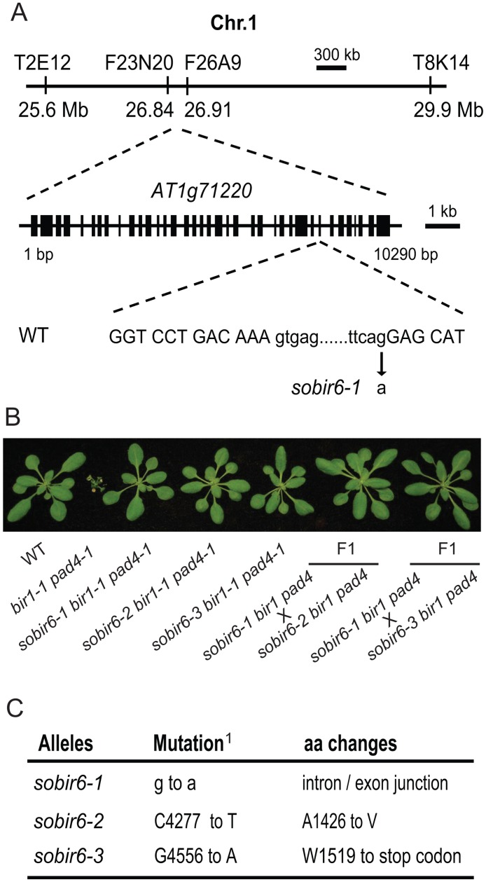
Fig 2. Map- based cloning of SOBIR6.
(A) Mapping of the sobir6–1 mutation. Positions of the mapping markers, the gene structure of SOBIR6 and the mutation site in sobir6–1 are shown. The exons are indicated with boxes and introns with lines. The mutation site is located at the junction between the 29th intron and 30th exon. The lower case letters represent nucleotides in the intron and the uppercase letters represent nucleotides in the exon. (B) Morphology of sobir6 bir1–1 pad4–1 alleles and representative F1 plants of indicated crosses for the complementation test. Plants were grown on soil at 23°C and photographed three weeks after planting. (C) Mutations identified in the sobir6 alleles. aa, amino acid. 1The positions of mutated nucleotide in the coding sequence are listed.

