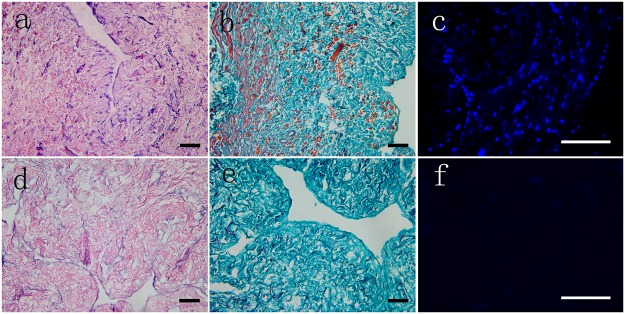
Fig 1. Histological analysis of fresh ureter tissue and its corresponding decellularized ECM via H&E, Masson’s trichrome and DAPI staining.

Cellular debris was obvious in the fresh tissue cross-sections (a, b) but had disappeared in the decellularized ECM (d, e). Intact and well-organized nuclei could be observed in the fresh tissue (c), whereas none could be seen in the decellularized ECM (f). Inset scale bar = 300 μm.
