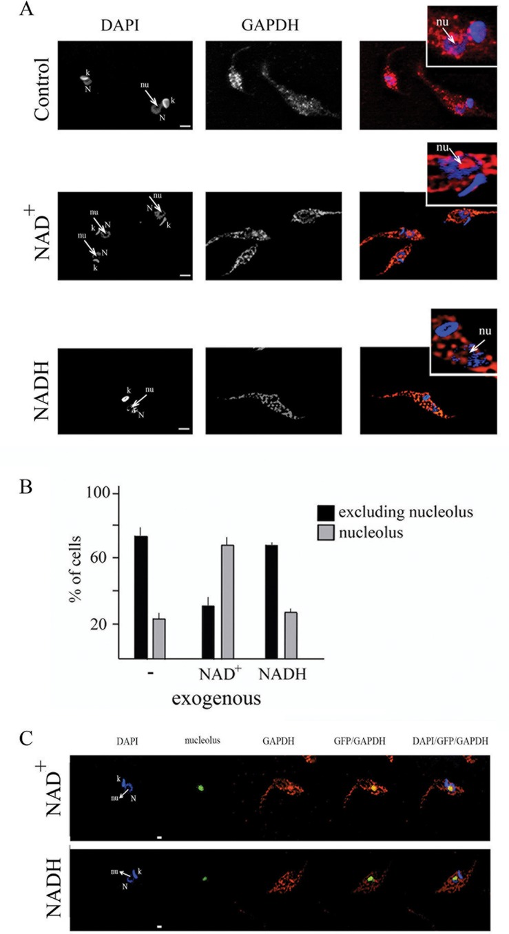
Fig 5. Exogenous NAD+ triggers translocation of GAPDH to nucleolus.
(A) T. cruzi epimastigote cells were maintained in the presence of NAD+ or NADH for 10 min, fixed, permeabilized and incubated with anti-GAPDH antibody (red). Cells were also stained with DAPI (blue). N- nucleus, k-kinetoplast and nu- nucleolus. Bars are 2μm. (B) Percentage of cells presenting GAPDH constrained at nucleolar space or dispersed through the nuclear space was quantified. Graph shows media and stand deviation of three independent experiments (n = 50 in each experiment). (C) T. cruzi epimastigote cells expressing GFP (green) that localizes in the nucleolus were maintained in the presence of NAD+ or NADH for 10 min, fixed, permeabilized and incubated with anti-GAPDH antibody (red). Cells were also stained with DAPI (blue). N- nucleus, k-kinetoplast and nu- nucleolus. Bars are 1μm.

