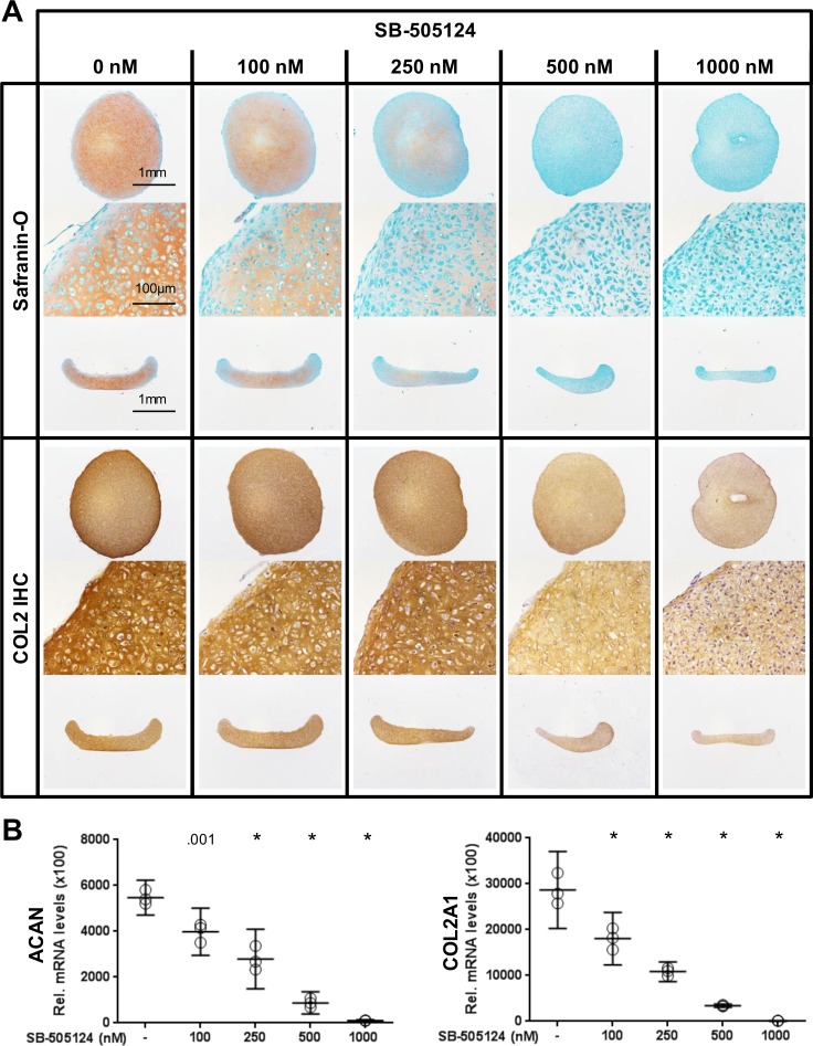
Fig 3. Blocking the TGF-β signaling pathway using the TβR1 kinase inhibitor SB-505124 in pellet cultures of P1 cells.
(A) The addition of SB-505124 to the pellet cultures of P1 cells abrogated autonomous cartilage-like tissue formation. Histological sections are shown from perpendicular axes (first and third row of Safranin-O and COL2 immunohistochemistry [IHC]). (B) The levels of transcripts coding for ACAN and COL2 decreased with increasing concentration of the inhibitor SB-505124. Data are shown as mean with the 95% confidence interval from one experiment with three separately analyzed cultures per group. The experiment was repeated once with cells from a different animal yielding similar results. Levels of transcripts of treated cultures were compared to control (0 nM) (ANOVA), indicated by exact P values or * P < .001.

