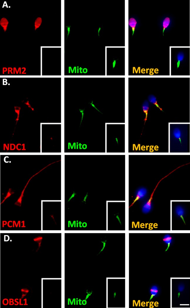
Fig 2. Localization of SEPT12 interactors in human-ejaculated spermatozoa.
Immunofluorescence detection of (A) PRM2, (B) NDC1, (C) PCM1, and (D) OBSL in human-ejaculated spermatozoa. Left panel: anti-SEPT12-interactor antibody (red). Middle panel: MitoTracker (Mito; green). Right panel: combination of the left and middle panels. Figure A–D. Staining with control IgG is shown in the right lower corner. More than 200 spermatozoa were evaluated at each antibody. Scale bar: 5 μm.

