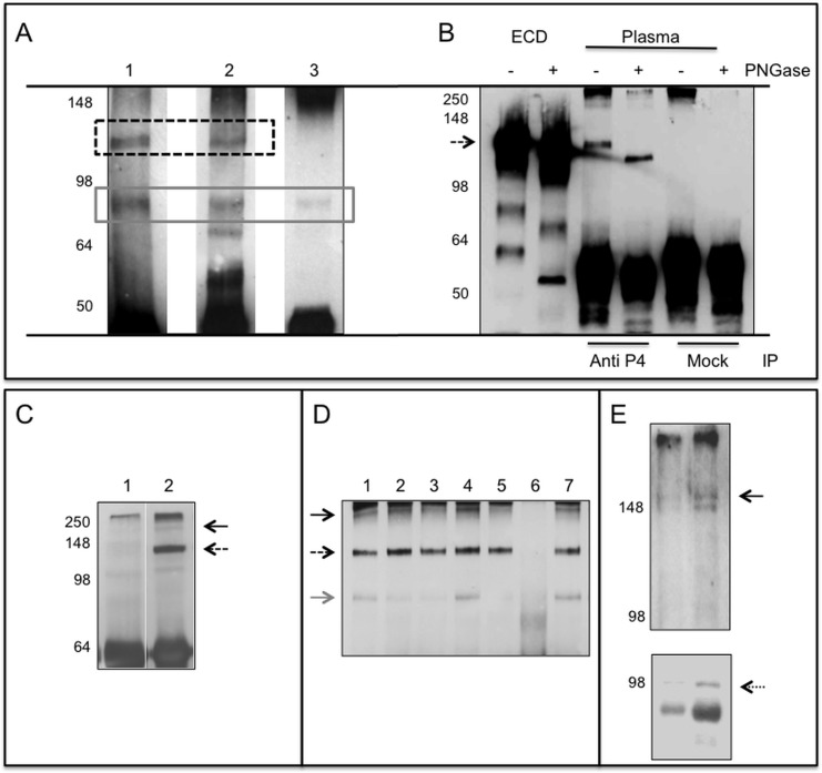
Fig 3. PTPRG plasmatic isoforms.
Panel A: Immunoblotting of human plasma samples immunoprecipitated (IP) with Rb anti-P4 and blotted against: Lane 1: Rb anti-P4, lane 2: mouse monoclonal R&D, lane 3: rabbit polyclonal Abcam. Dashed box: ∼120 kDa PTPRG isoform, gray solid box: ∼90 kDa isoform. Panel B: Immunoblotting with Rb anti-P4 of purified recombinant PTPRG-ECD-Fc lanes 1,2 and immunoprecipitated plasma samples with anti-P4 and RbIgG isotype control (Mock). Sample treated with the deglycosylating enzyme PNGase F are labeled with + sign. Dashed arrow: PTPRG ∼120 kDa isoform. Immunoblotting with Rb anti-P4 of anti-P4 IP human plasma (Panel C) and mouse serum (Panel D) of samples diluted 1:20 in PBS/Tx100 1%. Panel C: Lane 1: Immunoprecipitation with isotype control; Lane, 2: Rb anti-P4. Panel D: Lane 1–5 and 7: IP with Rb anti-P4. Lane 6: IP with RbIgG isotype control. Black arrow: full-length protein, dashed arrow: ∼120 kDa isoform, gray arrow: ∼90 kDa isoform. Left: molecular weight markers. Panel E: Plasma exosomes purified from two individuals were blotted with Rb-anti-P4 (upper panel) and anti ALIX rabbit antibody (lower panel, dotted arrow), an exosome marker. A band corresponding to full-length PTPRG is detectable in both samples (black arrow). Left: molecular weight marker.

