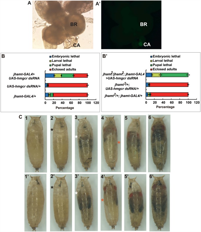
Fig 3. Reduction of hmgcr expression in the CA of jhamt mutant results in complete lethality.
(A and A’) The brain-RG complex in jhamt-GAL4>UAS-GFP. BR, brain; CA, corpus allatum. Observed under bright-field (A) or fluorescence (A’) using the same microscope. The CA cells expressing JHAMT were labeled with GFP. (B and B’) (B) Lethality of jhamt-GAL4>UAS-hmgcr dsRNA during the embryonic, larval, and pupal stages. jhamt-GAL4/+ and UAS-hmgcr dsRNA/+ were used as the controls. (B’) Lethality of jhamt 2/jhamt 2; jhamt-GAL4>UAS-hmgcr dsRNA during the embryonic, larval, and pupal stages. jhamt 2/+; jhamt-GAL4/+ and jhamt 2 /+; UAS-hmgcr dsRNA/+ were used as the controls. (C) Images of various pupal lethal phenotypes of jhamt 2 /jhamt 2; jhamt-GAL4>UAS-hmgcr dsRNA. (1–6) the abdominal sides; (1’-6’) the dorsal sides. The black asterisks point to empty portions of the pupae; the white asterisks, eye defects showing no pigmentation; the red asterisks, wing defects showing a unilateral wing loss.

