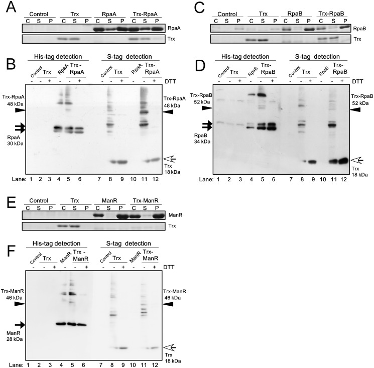
Fig 2. Examination of the interaction of RpaA, RpaB and ManR with Trx in E. coli cells.
Expression levels of (A) RpaA, (C) RpaB, (E) ManR and Trx in the control E. coli Origami2 strain (Control), the strain expressing only TrxMC35S (Trx), the strain expressing only a TF and the strain expressing both TrxMC35S and a TF were examined. The whole cell extract (C), the soluble fraction (S) and the insoluble pellet fraction (P) were separated by 15% SDS-PAGE and stained with CBB. The interaction of (B) RpaA, (D) RpaB and (F) ManR with Trx was examined by non-reducing 12% SDS-PAGE and immunoblot analysis of soluble proteins from each strain. TFs and Trx were detected using a His-tag antibody and S-protein, respectively. ± indicates with or without 100 mM DTT treatment. Black arrow, white arrow and arrow head indicate the TF monomer, the Trx monomer, and the Trx-TF complex, respectively.

