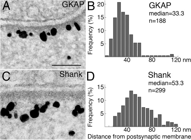
Fig 4. Distributions of label for GKAP and Shank are different at the PSD complex.
Higher magnification EM micrographs show that under basal conditions, label for GKAP is in a narrow band closer to the postsynaptic membrane (A), while label for Shank is located in a wider band in the PSD complex (C). Scale bar = 0.1 μm. Distance measurements of label are plotted in histograms (B for GKAP and D for Shank) showing significant difference in distribution (P< 0.0001, Wilcoxon rank-sum test) with a 20 nm difference in median values.

