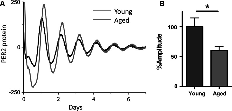
Fig. 3.
Age related decline in circadian amplitude in cartilage tissue explants. Adapted from Gossan et al. (2013): a representative trace of PER2::luc bioluminescence rhythms from xiphoid cartilage of young (2–4 months) and aged circadian reporter mice; b quantification of circadian amplitude showing a 40 % reduction in rhythm strength in aged (20–24 months) cartilage, mean ± SEM, n = 8 young, 11 aged, t test, p < 0.05

