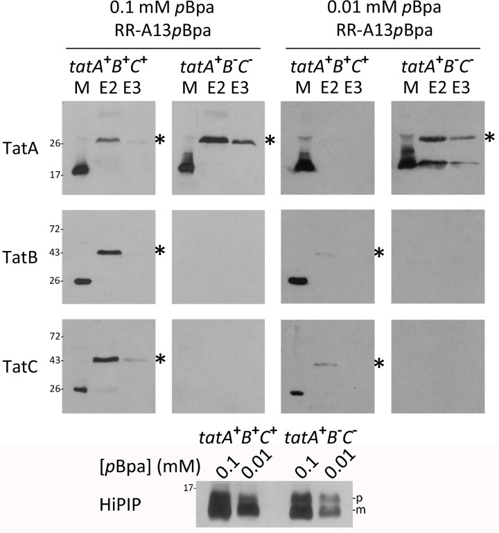
Fig 2. The TatA/Tat signal peptide contact does not require the TatBC components.
Comparison of HiPIP-TatA cross-links in the presence/absence of TatBC using pBpa at position A13. Two pBpa concentrations (0.1 mM and 0.01 mM) in the growth medium were used to analyze effects of lowered HiPIP concentration. tatA + B + C +: BW25113/pRK-tatABC/pEvol-pBpF /pEXH5tac-H6-A13pBpa; tatA + B - C -: JBdBC/pRK-tatA/pEvol-pBpF /pEXH5tac-H6-A13pBpa. The bottom blot monitors the decrease of HiPIP levels by reduction of pBpa concentration in the medium. HiPIP detection in crude extracts. Significant HiPIP degradation to mature size is due to unspecific proteolysis of the signal peptide. See Fig. 1 for further details.

