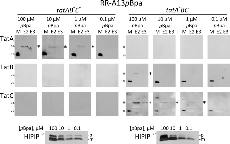
Fig 3. Substrate cross-links to TatA and TatC have comparable detection limits when substrate levels are gradually decreased.
Detection of cross-links of RR-HiPIP with pBpa at position A13 (RR-A13pBpa) either to wild-type level non-recombinant TatA in the absence of TatBC (left section: tatAB - C -, strain JBdBC) or to wild-type level non-recombinant TatBC in the absence of TatA (right section: tatA - BC; strain JARV16). Note that TatA and TatC cross-links are detectable at lowered substrate concentrations as achieved with 1 μM pBpa in the medium. TatB cross-links are depleted below detectability already with 10 μM pBpa, whereas TatA and TatC cross-links both become non-detectable with 0.1 μM pBpa. Bottom blots: SDS-PAGE-Western blot detection of HiPIP in extracts of cells grown in the presence of indicated pBpa concentrations. The bottom blots monitor the decrease of HiPIP levels by reduction of pBpa concentration in the medium, as described in Fig. 2. p, precursor of HiPIP; m, mature form of HiPIP. See Fig. 1 for further details.

