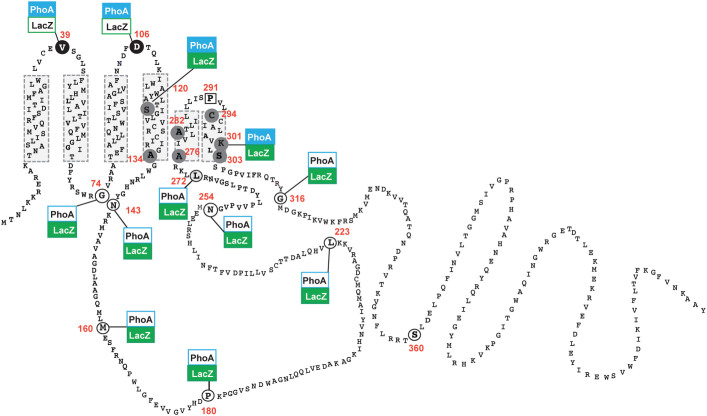
Figure 3. Refined topological model of the E. coli WcaJ based on reporter fusions and substituted cysteine labelling with PEG-Mal.
Blue coloured boxes indicate positive PhoA results and green coloured boxes indicate positive LacZ results. White coloured boxes indicate negative reporter fusion results. Residues indicated in circles were replaced by cysteine in the WcaJCysless version of the protein. Residues that were labelled with PEG-Mal denoting a periplasmic location are depicted with black background. Residues indicated in grey are those only detected by PEG-Mal after SDS solubilization, indicating they are buried in the membrane bilayer. Residues exposed to the cytosol are depicted in white background. The square indicates the position of the conserved P291, which could not be labelled with PEG-Mal under any of the treatments employed including SDS solubilization of membrane fractions.

