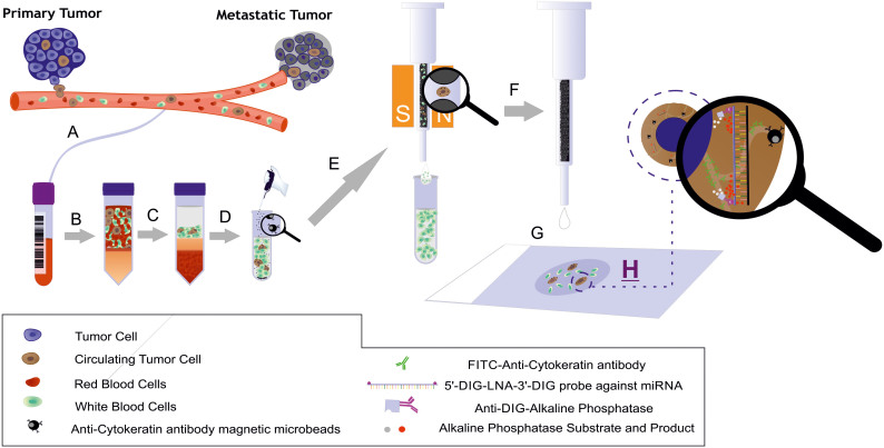
Figure 1. Schematic illustration of the MishCTC method for simultaneous miRNA and CK detection via immunocytochemistry.
(A) Recovery of peripheral blood into an EDTA tube; (B) blood transfer into a density-gradient centrifuge tube; (C) centrifugation at 700 × g for 30 min; (D) recovery of the interphase layer, which contains mononuclear and tumor cells, and immunomagnetic labeling with magnetic microbeads conjugated to an anti-CK antibody; (E) magnetic cell separation using a MiniMACS separator and a pre-filled separation column; (F) elution of retained cells; (G) application of the cells to a polylysine glass slide using CytoSpin centrifugation; and (H) MishCTC detection of miRNA and CK. Mr. Juan M. Agudo helped to prepare this artwork.

