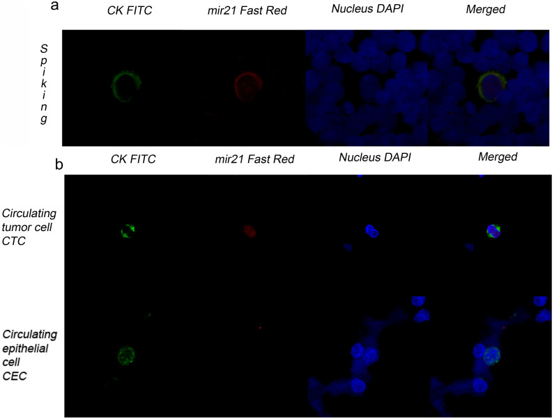
Figure 2. Image galleries obtained with the MishCTC method.
(a) CK and miRNA-21 expression in an MDA-MB468 cell that was spiked into a blood sample from a healthy volunteer. Detection of cytokeratin-positive (CK+) cells (green channel), miRNA-21-positive cells (red channel) and nuclei (blue channel). Epithelial cells were identified in a leukocyte population that did not express miRNA-21. (b) CK and miRNA-21 expression in a CTC from a patient with metastatic lung cancer. All the CTCs that were found within this set of patients were both CK- and miRNA-positive (upper panel). CK expression in a circulating epithelial cell from a cancer-free patient undergoing a nephrectomy. CK protein expression (green) was detected by immunofluorescence, but miRNA-21 could not be detected by in situ hybridization (lower panel).

