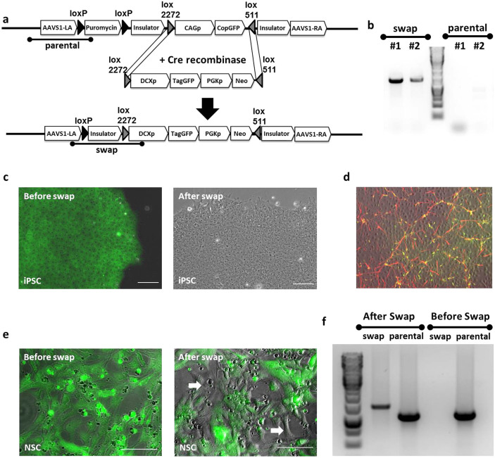
Figure 2. Rapid exchanging of reporter cassettes in safe harbors in iPSC and progenitor cells using a master cell line strategy.
(a) The experimental strategy of quick and efficient generation of a reporter line via RMCE strategy. The master line, AAVS1-copGFP, was co-transfected with the Cre expression vector and the targeting plasmid DCXp-TagGFP carrying a lox2272-DCXpromoter-TagGFP-PGK-Neo-lox511 cassette for RMCE with the master line. Cells with successfully targeted recombination were neomycin resistant and expressed TagGFP under the endogenous promoter of DCX instead of the constitutive CAG promoter which is seen in the master line. Testing primers, “swap” (indicating the successful RMCE event) and “parental” (detecting the parental gene which has no swap event) were also illustrated. Solid black triangles represent the LoxP sites and triangles filled with diagonal lines represent Lox sites for RMCE. (b) PCR verification of the selected non-fluorescent colonies. No “parental” PCR products were detected in any of the selected colonies. (c) After Neomycin selection, colonies with no copGFP signal were selected under fluorescent microscope. Overlaying fluorescence channel with the bright field, the cells before swap were all green fluorescent (left). After correct swap, the CAG promoter driving copGFP was replaced by DCX promoter driving TagGFP whose expression is off at the iPSC stage, and cells were no longer fluorescent (right). (d) Neuronal differentiation was induced on iPSC selected above and immunostaining showed positive co-localization of DCX antibody (red) and TagGFP (green), suggesting that the TagGFP is only expressed when the DCX gene is turned on. (e) RMCE strategy was also tested in the progenitor stage (NSC). AAVS1-copGFP master line NSC were co-transfected with Cre expressing vector and the targeting plasmid DCXp-TagGFP as described in (a). Before transfection, all AAVS1-copGFP NSC were green fluorescent (left). After 5 days post transfection, cells lost green fluorescence were detected under microscope (spots pointed by arrows in the right graph). Images shown here are bright field superimposed with fluorescence channel. (f) PCR verification of successful swapping event happened in NSC. Only “parental” PCR band was detectable in NSC before transfection. After RMCE, both “swap” and “parental” PCR products were detected in the mixed culture. Scale bar is 100 μm.

