
Figure 3.
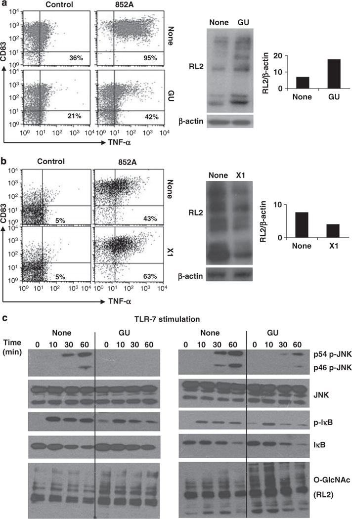
Effect of changing O-GlcNAc levels on toll-like receptor (TLR)-7-signaling in chronic lymphocytic leukemia (CLL) cells. (a) CLL cells from patient 116 were cultured overnight, alone or in GlcNAc and Uridine (GU), before stimulation with the TLR-7 agonist, 852A (0.1 μg/ml). Immunoblotting with RL2 antibodies confirmed the increase in O-GlcNAcylated protein levels (middle panels), which was quantified (relative to β-actin levels) by densitometry (right panel). Surface expression of CD83 and tumor necrosis factor (TNF)-α (to reflect TLR-7-signalling10) were then measured by flow cytometry after 4h. Numbers in the right upper quadrants are the percentages of CLL cells that express membrane TNF-α (mTNF-α). (b) CLL cells from patient 164 were cultured for 24h alone or with the OGT-inhibitor, X1 (50 μM), to decrease O-GlcNAcylation (confirmed by immunoblotting with RL2 antibodies and densitometry (middle and right panels) before stimulation with 852A. Similar increases in TNF-α production were seen with ten other samples. (c) CLL cells from patient 80 (left panel) and patient 125 (right panel) were cultured overnight in the presence or absence of GU and then stimulated with 852A. At the indicated times, protein extracts were collected and expression of phosphorylated and total c-Jun N-terminal kinases (JNK) and IκB, as well as RL2, to confirm the increased levels of O-GlcNAcylated proteins, were examined by immunoblotting. Similar results were seen with two other samples.