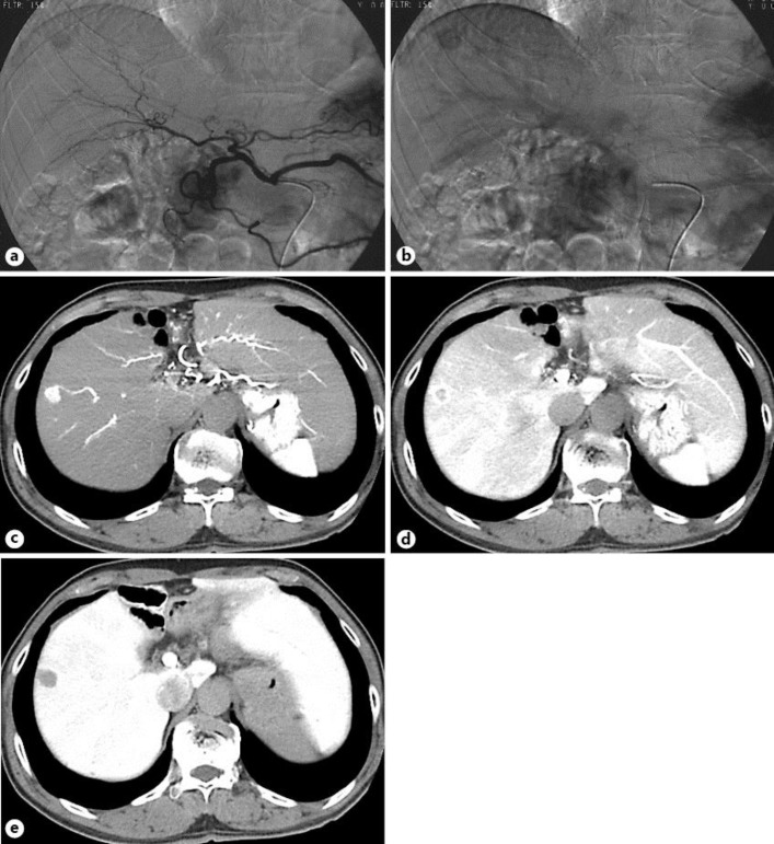
Fig. 2.
Hepatic angiography in the early phase (a) and late phase (b) revealed a tumor stain in the anterior superior arteries. The mass in the liver (S8) was described as an enhanced lesion on CT during hepatic arteriography (c), a corona-like enhanced lesion on CT in the portal-venous phase (d), and a low-density lesion on CT during arterial portography (e).

