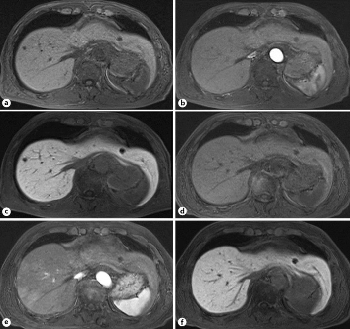
Fig. 3.
MRI revealed a 6-mm low-signal intensity mass in the liver (S8) in T1-weighted image (a) 2 months after the initial diagnosis. The gadolinium ethoxybenzyl diethylenetriamine pentaacetic acid-enhanced MRI disclosed no mass in the arterial phase (b) and a 6-mm low-signal intensity mass in the hepatobiliary phase (c). MRI showed no mass in the liver (S8) in the pre-enhanced phase of T1-weighted image (d), arterial phase (e), and hepatobiliary phase (f) 6 months after the initial diagnosis.

