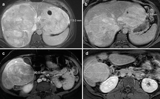
Fig. 1.

Post-contrast (gadoxetate disodium) MRI of a patient with GSD I with HCAs. (a) Fat-saturated T1-weighted post-contrast axial MR image of the superior liver obtained after intravenous bolus injection of gadoxetate disodium demonstrates multiple large lesions in the liver consistent with HCAs, two of the lesions are measured. (b) Fat-saturated T1-weighted post-contrast axial MR image of the superior liver obtained after intravenous bolus injection of gadoxetate disodium in the same patient 4 years later demonstrates decrease in size of the multiple HCAs, two of which are measured for comparison. (c) Fat-saturated T1-weighted post-contrast axial MR image of the inferior liver obtained after intravenous bolus injection of gadoxetate disodium in the same patient at the same time as the image in (a) demonstrates a large HCA involving the inferior right hepatic lobe (lesion is measured). (d) Fat-saturated T1-weighted post-contrast axial MR image of the inferior liver obtained after intravenous bolus injection of gadoxetate disodium in the same patient four years later, at the same time as the image in (b), demonstrates significant decrease in size of the HCA, measured for comparison
