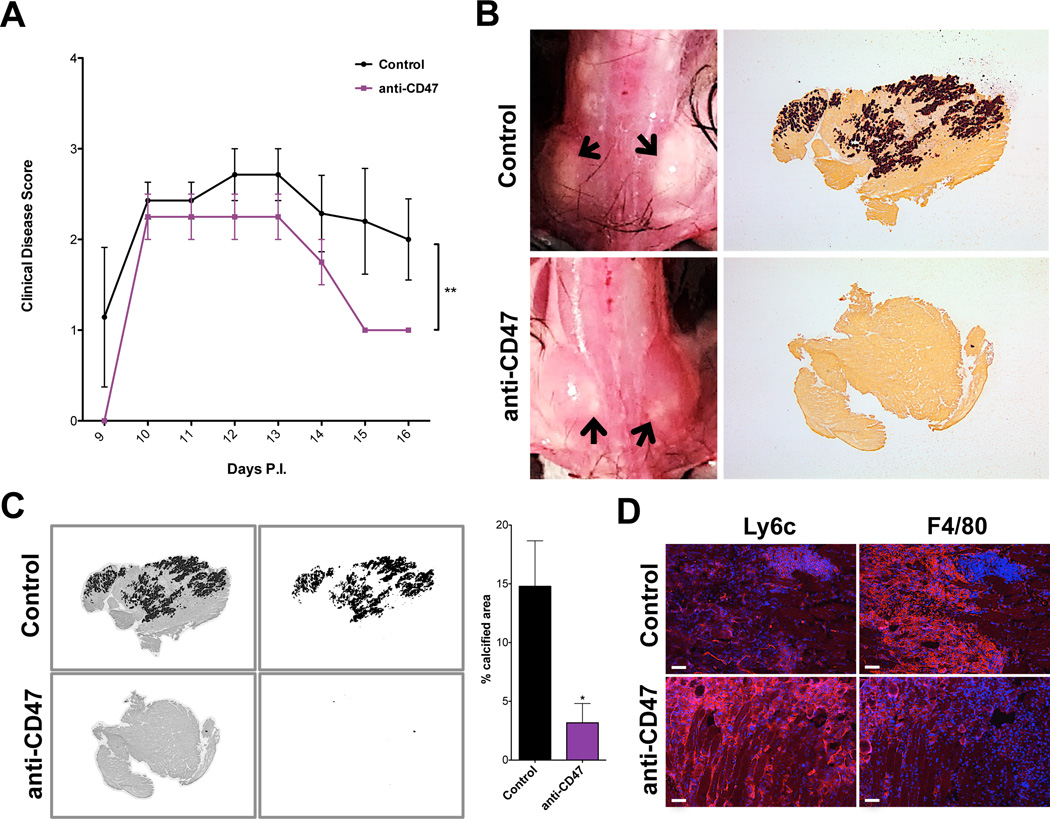
Figure 5. Impact of CD47 neutralization on disease severity.
WT BL/6 mice were infected with TMEV and injected with control or CD47-neutralizing antibodies from 5–13 days P.I. (A) Mean clinical disease scores (+/− SEM) of mice according to the following scale: 1) hunched posture and ruffled fur; 2) swollen muscles/abnormal gait; 3) 1 limb immobilized; 4) 2+ limbs immobilized; 5) death. (B) Muscle calcification observed upon dissection at 16 days P.I. Left panels: representative images of muscle calcification from uninjected and anti-CD47-injected mice; arrows indicate calcified areas. Right panels: representative alizarin red stainings of muscle from uninjected and anti-CD47-injected mice (2× magnification). (C) Percent calcified area of muscle was measured by converting brightfield images of alizarin red stains into 8bit black and white files (left images), measuring the area of dark staining (right images) and measuring the whole tissue area. Representative pictures shown of IgG-injected control and anti-CD47 Ab-injected animals; graph depicts percent calcified area. Uninjected and IgG-injected controls were pooled (n=7); n=3 for CD47-neutralized animals. (D) Frozen muscle from control and CD47-neutralized animals at 16 days P.I. was labeled with antibodies against Ly6c (left) and F4/80 (right) in red and stained with DAPI (blue). Representative images shown of untreated control and anti-CD47-injected mice; control n=7, anti-CD47 n=4; scale bars depict 100µm; experiment performed in duplicate.

