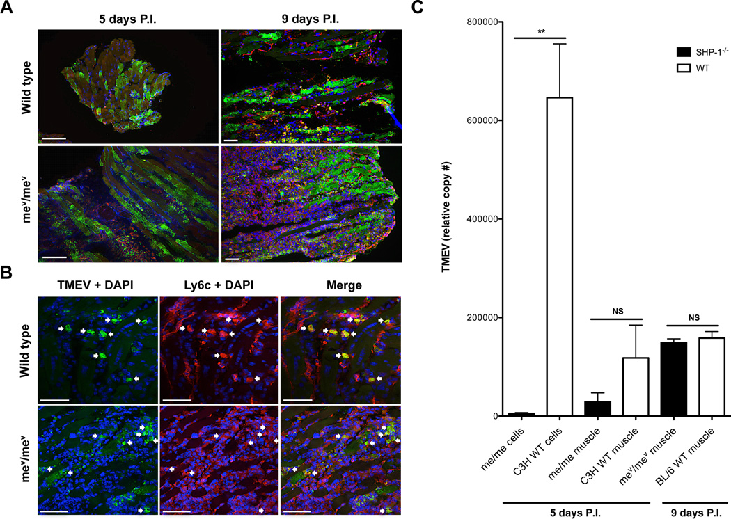
Figure 8. TMEV replication in WT and SHP-1−/− muscle.
(A) mev/mev mice and wild type littermates were sacrificed at 5 or 9 days P.I. and muscle sections were labeled with TMEV (green) and stained with DAPI (blue); n=3. Sections from animals sacrificed at 5 days P.I. were labeled with antibodies against F4/80 (red), scale bars represent 50µm. Sections from animals sacrificed at 9 days P.I. were labeled with antibodies against Ly6c (red); scale bars represent 100µm. (B) mev/mev mice and wild type littermates were sacrificed at 9 days P.I. and muscle sections were labeled with antibodies against Ly6c (red), TMEV (green) and stained with DAPI (blue); arrows suggest infected Ly6c+ cells. Scale bars represent 50µm, n=3. (C) TMEV RNA levels were quantified from CD45+CD11b+Ly6c+ cells sorted from C3H me/me and WT littermates at 5 days P.I. (me/me and C3H WT cells), and homogenized muscle of C3H me/me and WT littermates at 5 days P.I. (me/me and C3H WT muscle) and BL/6 mev/mev and WT littermates at 9 days P.I. (mev/mev and BL/6 WT muscle). N=3; C3H me/me and WT muscle p=0.1329; BL/6 mev/mev and WT muscle p=0.2890.

