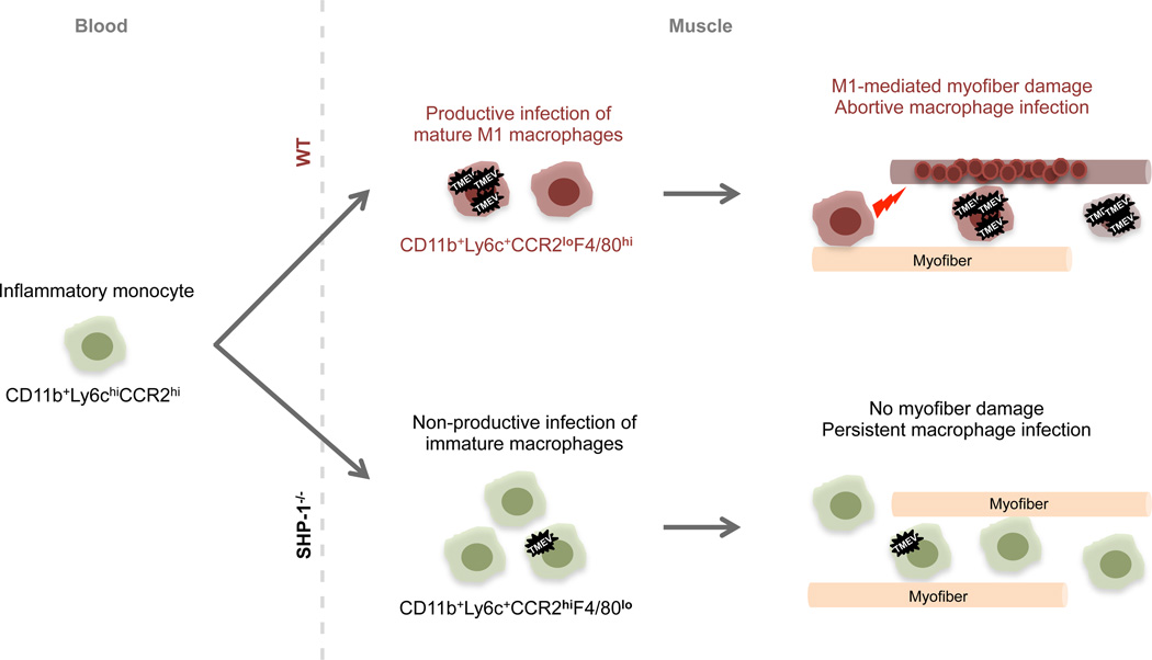
Figure 9. Proposed mechanism of SHP-1-mediated TMEV pathogenesis.
Following TMEV infection of skeletal muscle, inflammatory monocytes (green) infiltrate skeletal muscle in WT and SHP-1−/− mice. Top: in WT mice, monocytes differentiate into M1-like macrophages (red), supporting TMEV replication and contributing to muscle fiber damage and subsequent calcification (red circles) likely via proinflammatory cytokines (red lightening bolt). Following active replication TMEV produces an abortive infection, inducing apoptosis of macrophages and limiting the numbers of infiltrating cells. In the absence of SHP-1 (bottom), abnormally high numbers of muscle-infiltrating monocytes remain immature (green), preventing efficient replication of TMEV. These immature monocyte-like cells do not undergo apoptosis and do not produce disease pathology in skeletal muscle.

