Figure 2.

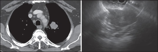
Left – Left upper-lobe mass, right – Endoscopic ultrasound image of fine-needle aspiration through a narrow window superior and posterior to the aortic arch

Left – Left upper-lobe mass, right – Endoscopic ultrasound image of fine-needle aspiration through a narrow window superior and posterior to the aortic arch