
Figure 6.
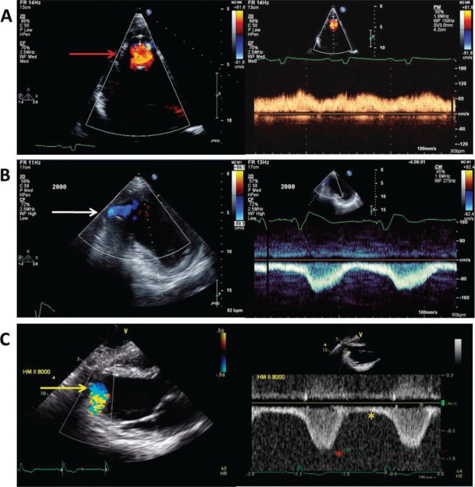
Normal continuous left ventricular assist device apical inflow cannula color and spectral Doppler. (A) Standard 4-chamber apical view illustration of nonturbulent, nonaliasing apical cannula inflow with color Doppler (red arrow) and slightly pulsatile, low peak velocity (< 1.5 m/s) continuous flow directed towards the apical transducer. (B) “Off axis” 2-chamber apical view of an inferiorly positioned apical inflow cannula (white arrow) with normal flow characteristics (slightly pulsatile, low peak velocity) similar to panel A with the exception of continuous flow directed away from the apical transducer. Red asterisk shows peak systolic apical inflow velocity; yellow asterisk shows peak diastolic apical inflow velocity. (C) Parasternal long-axis view of normal apical cannula inflow color Doppler (yellow arrow) and pulse-wave Doppler flow characteristics with continuous flow directed away from the apical transducer similar to panel B. Reprinted from Estep et al. with permission of the publisher. Copyright ©2010 Elsevier.14