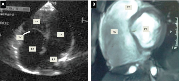
Figure 2.

Images showing the A) transthoracic echocardiogram of the low implantation of the TV and RV atrialization was visualized in a 4-chamber view. B) Vectographic image using MR showed extensive RA dilatation (53 × 103 mm) with atrialization of the RV inflow tract and showed the TV with abnormal apical implantation of the septal leaflet (31 mm). MR - magnetic resonance, RA - right atrium, TV - tricuspid valve, LA - left atrium, RV - right ventricle, LV - left ventricle.
