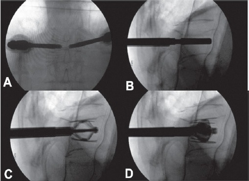
Figure 3.

Intraoperative radiographs demonstrating the main steps of the technique. The anterior-posterior (A) and lateral (B) view demonstrate bilateral percutaneous transpedicular placement of the implant. The implant is expanded (C) and after adequate restoration, the cement is injected under fluoroscopic monitoring.
