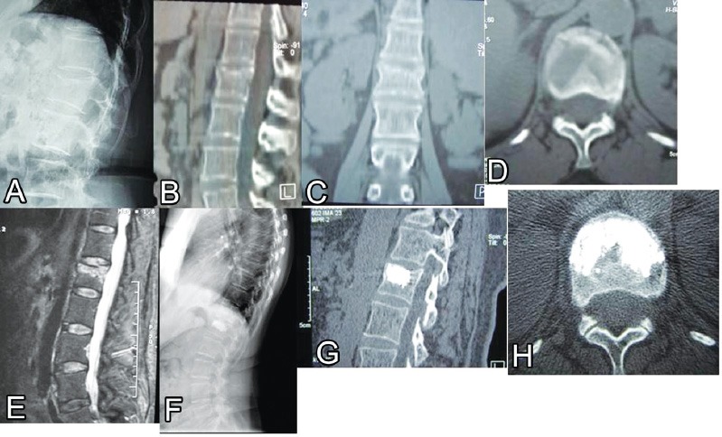
Figure 4.

A case illustration of a 42-year-old male presented after a trivial fall with severe lower back pain and normal neurological exam (visual analogue scale 9). Lateral radiograph (A) demonstrated vertebral compression fractures (VCF) (type A1.3). Sagittal B), coronal C), and axial D) CT scans demonstrate fracture of the upper endplate and kyphotic deformity. A T2-WI sagittal MRI scan E) demonstrated high signal intensity (edema) at the fracture site. One-year follow up lateral radiograph F) showing adequate fusion of the VCF was demonstrated on sagittal G) and axial H) CT scans.
