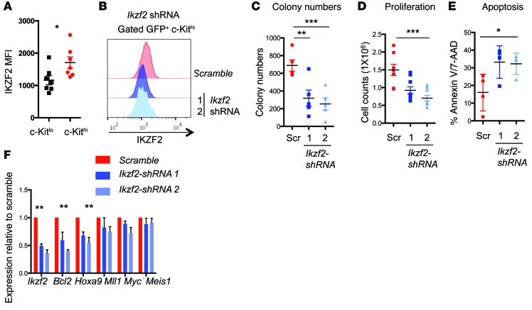
Figure 6. Ikzf2 contributes to the survival of myeloid leukemia cells.
(A) Median fluorescence intensity of IKZF2 in the c-Kithi and c-Kitlo MLL-AF9 leukemic cells (n = 7, combined from 2 independent experiments). (B) Representative flow cytometric plot for intracellular staining of IKZF2 showing knockdown in c-Kithi cells derived from Msi2fl/fl leukemia cells transduced with scramble and 2 Ikzf2 shRNAs (1 and 2) from 3 experiments. (C) Colony formation assay of MLL-AF9 leukemic cells transduced with scramble or Ikzf2 shRNAs, same as in B, plated in methylcellulose and counted after 5 days; n = 5. (D) Proliferation at 6 days post-transduction; n = 4. (E) Apoptosis at 6 days post-transduction; n = 4. (F) Quantitative PCR of MLL-related genes in MLL-AF9 leukemic cells transduced with scramble or Ikzf2 shRNAs. Results are means and SEM of n = 4 for scramble and Ikzf2 shRNA2 and n = 3 for Ikzf2 shRNA1. *P < 0.05, **P < 0.01, ***P < 0.001, unpaired Student’s t test for A to F.

