
Figure 1. ENS morphogenesis.
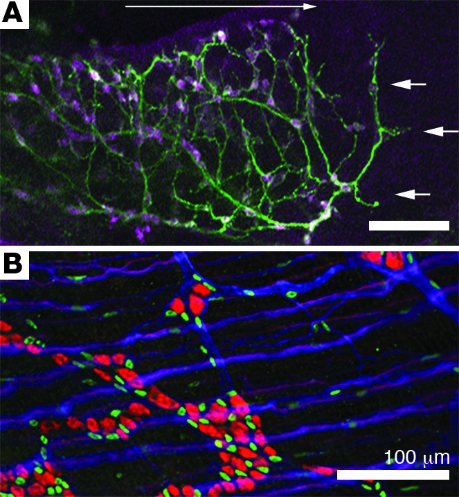
(A) Vagal ENCDCs migrate in a rostral to caudal direction through fetal bowel (long white arrow). At E12.5 ENS precursors have migrated halfway through the fetal colon. ANNA-1 antibody binds HuC/D antigen and identifies enteric neurons (magenta), while TuJ1 binds neuron-specific β-III tubulin and labels neurites (green). ENCDCs migrate in chains though the bowel, but during the period of migration some precursors differentiate into neurons and extend neurites, including at the migration wavefront (white arrows). (B) Adult small bowel myenteric plexus, indicated by ANNA-1 antibody (red, neurons), SOX10 antibody (green, enteric glia), and TuJ1 antibody (blue), demonstrates clusters of neurons and glia in mature ganglia as well as many neurites within and between ganglia. Scale bars: 100 microns.