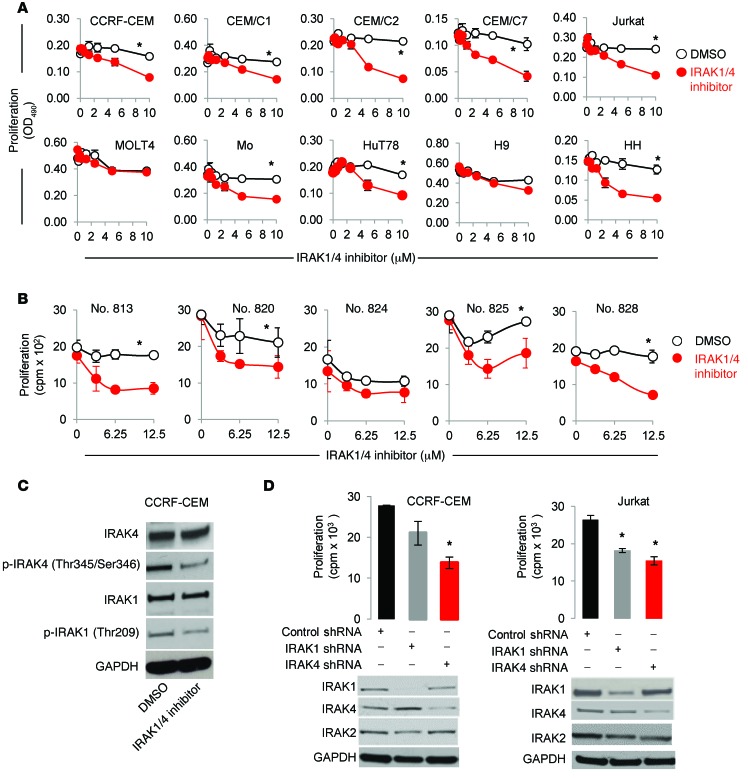
Figure 2. IRAK1/4 signaling inhibition impedes T-ALL proliferation.
(A) IRAK4/1 inhibitor effects on the proliferation of various malignant T cell lines. Cells were cultured in the presence of IRAK1/4 inhibitor (0–10 μM) or control (DMSO), and proliferation was measured 72 hours later. Data are representative of 3 independent experiments, and the average OD490 (± SD) of triplicate readings is shown. (B) Patient T-ALL cells were cultured in the presence of control (DMSO) or IRAK1/4 inhibitor (0–12.5 μM), and proliferation was determined by measuring 3[H]thymidine incorporation. Data represent the average of triplicate readings (± SD) and are representative of 2 independent experiments, each yielding identical trends. *P <0.05 by Student’s t test (A and B). (C) Levels of the indicated proteins were examined in CCRF-CEM T-ALL cells after treatment with IRAK1/4 inhibitor (2.5 μM) or DMSO for 48 hours. (D) T-ALL cells were transiently transfected with the indicated plasmids. Transfected cells were sorted by flow cytometry according to GFP expression, and proliferation of CCRF-CEM and Jurkat cells was determined according to 3[H]thymidine uptake (top panels). Average cpm (± SD) of triplicate readings is shown. *P < 0.05 versus control shRNA by Student’s t test. Knockdown efficiency was examined by Western blotting. Data are representative of 3 independent experiments.

