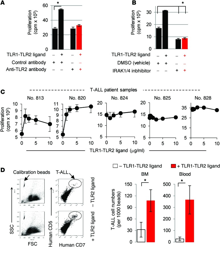
Figure 3. TLR1-TLR2 engagement can augment T-ALL proliferation.
(A) CCRF-CEM cells were treated with Pam3CysK4 (2.5 μg/ml) or control (PBS) in the presence or absence of either 5 mg/ml neutralizing anti-TLR2 antibody or isotype control antibody. After 48 hours, proliferation was determined by 3[H]thymidine incorporation. (B) CCRF-CEM cells were cultured with DMSO or IRAK1/4 inhibitor (1.5 μM) in the presence or absence of Pam3CysK4 (2.5 μg/ml), and after 48 hours, proliferation was determined by 3[H]thymidine incorporation. Data in A and B are representative of 3 independent experiments, and the average cpm (± SD) of triplicate readings is shown. *P < 0.05 by Student’s t test. (C) Patient T-ALL samples were treated with the indicated concentrations of Pam3CysK4 for 48 hours. Proliferation was determined by 3[H]thymidine incorporation, and data are representative of 2 experiments. (D) CCRF-CEM cells (3 × 10⁶) were i.v. injected into NSG mice (n = 5). Seven day later, the mice were injected with Pam3CysK4 (2 mg/kg) or control (PBS), and the number of T-ALL cells in circulation and BM was determined on day 14 by staining cells with anti–human CD7 and CD5 antibodies and analyzed by flow cytometry. Cell counts were normalized by adding an equal volume of calibration beads to 50 μl blood and setting the instrument gates to count a constant number of beads. Representative dot plots (left panel) and absolute numbers in BM and blood (right panel). Data represent the average cell counts (± SD) in 5 mice and are representative of 2 independent experiments. *P < 0.05 by Student’s t test. FSC, forward scatter; SSC, side scatter.

