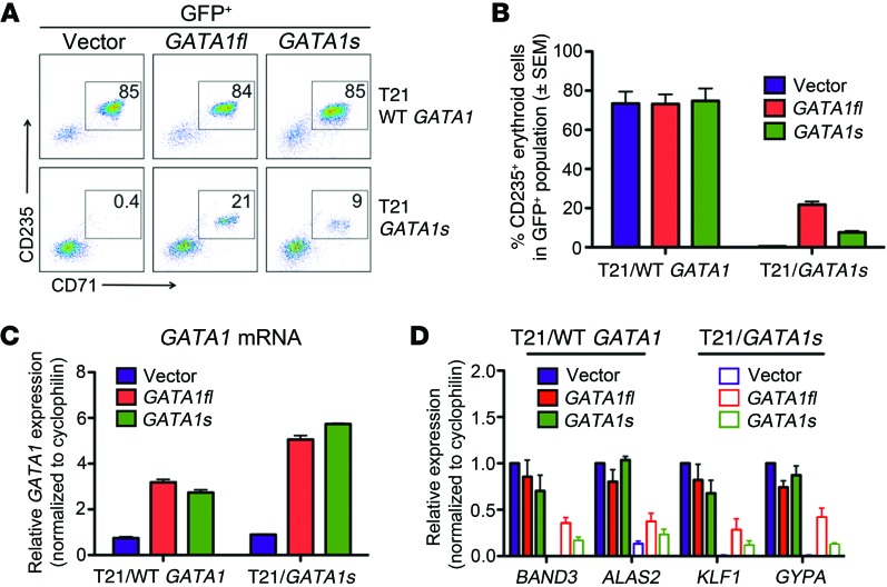
Figure 3. Dose-dependent restoration of erythropoiesis with enforced expression of full‑length GATA1 and truncated GATA1s.
T21/WT GATA1 and T21/GATA1s iPSC–derived CD43+CD41+CD235+ progenitors were transduced with lentivirus containing vector alone or encoding GATA1fl or GATA1s and cultured with EPO and SCF (n = 4 replicates). (A) Representative flow-cytometric analysis after 6 days of culture. Numbers denote percentage of total cells in the indicated gate. (B) Average percentage of CD235+ erythroblasts in transduced (GFP+) cells after 6 days of culture. (C) PCR showing relative GATA1 mRNA expression 2 days after lentiviral infection with control vector, GATA1fl, or GATA1s. The genotype of progenitors is shown on the x axis. (D) Expression of selected erythroid mRNAs in flow-cytometry–purified CD235+ infected (GFP+) cells.

