
Figure 3.
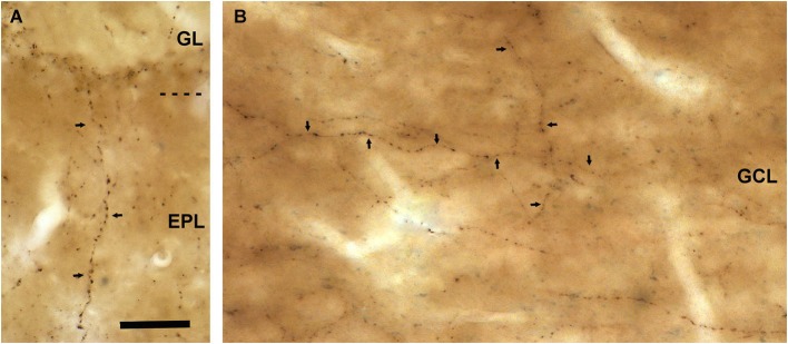
VAChT-staining in the external plexiform layer and in the inframitral region under light microcopy. (A) A varicose axon containing VAChT (arrows) crosses the external plexiform layer (EPL) perpendicularly to the bulbar lamination. The glomerular layer (GL) is shown in the upper side of the micrograph. (B) VAChT-containing fibers (arrows) running throughout the superficial portion of the granule cell layer (GCL). Note that some fibers run parallel and others perpendicularly to the bulbar lamination. Scale bar: 30 μm.