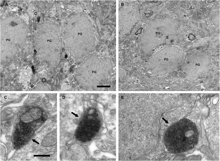
Figure 4.
Perisomatic innervation of periglomerular cells by VAChT-containing boutons under electron microscopy. (A,B) Low-magnification views of the periglomerular region of the glomerular layer showing the somata of periglomerular cells (PG) surrounded by VAChT-containing boutons. The squared boutons are shown at higher magnification in the panels (C–E). (C–E) Synaptic contacts (arrows) from the VAChT-containing boutons on the somata of periglomerular cells. Scale bars: 2 μm in (A) and (B); 200 nm in (C), (D) and (E).

