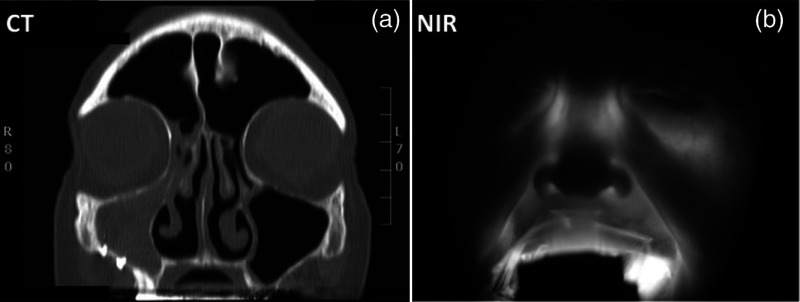
Fig. 1.
Example of near-infrared (NIR) optical imaging in the maxillary sinus. The patient presents a blocked right sinus as indicated in the computed tomography (CT) image slice (a). The patient’s left sinus is clear as shown by the thin white lining in the void region (black). The patient’s right sinus is completely filled (i.e., high opacity). The corresponding NIR image (b) mirrors the opacity of the CT image. The light source, composed of 850 nm LEDs, was placed inside the mouth to transilluminate the sinus. The bright aura around the mouth is due to source light leakage through the mouth area.

