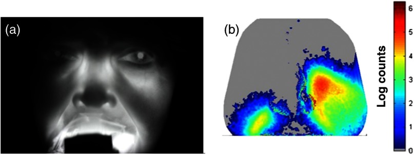
Fig. 7.
Computer simulation using 3-D CT scan and Monte Carlo model compared to experiment for a case of advanced sinusitis. The CT image for patient #2 is provided in Fig. 1 and reveals that the right maxillary sinus is completely blocked. The high opacity of the sinus is evident in the NIR image of the patient (a). Using the patient’s 3-D CT scan slices, we simulate what the clinical image should look like in (b). The main features of the image (low opacity on left, high opacity on right) are clearly visible. In this case, the illumination of the region near the ethmoid sinuses is not visible in the simulation.

