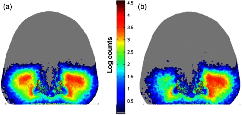
Fig. 9.
Simulations showing progression of sinus disease. (a) The simulation of the case corresponding to the clinical image of patient #3 (Fig. 8). (b) A simulation of further disease progression where we increased mucosal thickening by an additional 3 mm. Note that the trend of asymmetry continues and becomes significantly visible with increased mucosal thickening. Light attenuation increases as a result of the reduced air volume in the sinus due to mucosal thickening.

