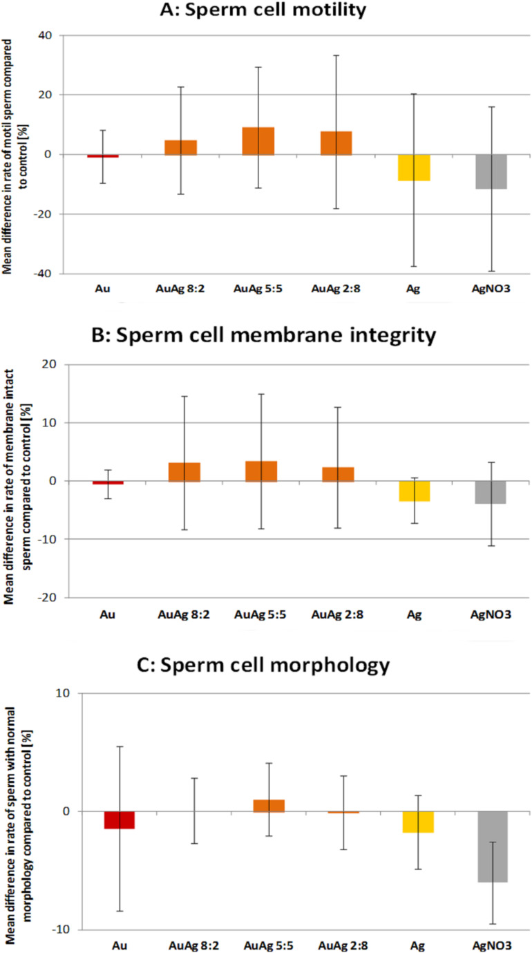
Figure 4.
Sperm viability parameters after co-incubation of sperm for 2 h at 37 °C with various nanoparticle types and a silver nitrate control. Nanoparticle concentration was 10 µg/mL. (A) Motility assessed with Computer Assissted Sperm Analysis, (B) Membrane integrity assessed with propidium iodide stain and flow cytometer, (C) morphology assessed with phase contrast microscope and evaluation of 200 sperm cells per group per day. Shown are percentage of spermatozoa, which differ compared to the control [values are mean ± SD]. Reproduced with permission from [50]. Copyright 2014 Royal Society of Chemistry.

