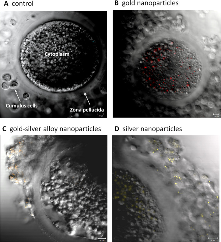
Figure 6.
Representative laser scanning microscope images of porcine cumulus–oocyte complexes after 46 h co-incubation during in vitro maturation. (A) Negative control; (B) gold nanoparticles; (C) gold–silver alloy nanoparticles; (D) silver nanoparticles; bars = 10 micrometer. Reproduced with permission from [50]. Copyright 2014 Royal Society of Chemistry.

