
Figure 5.
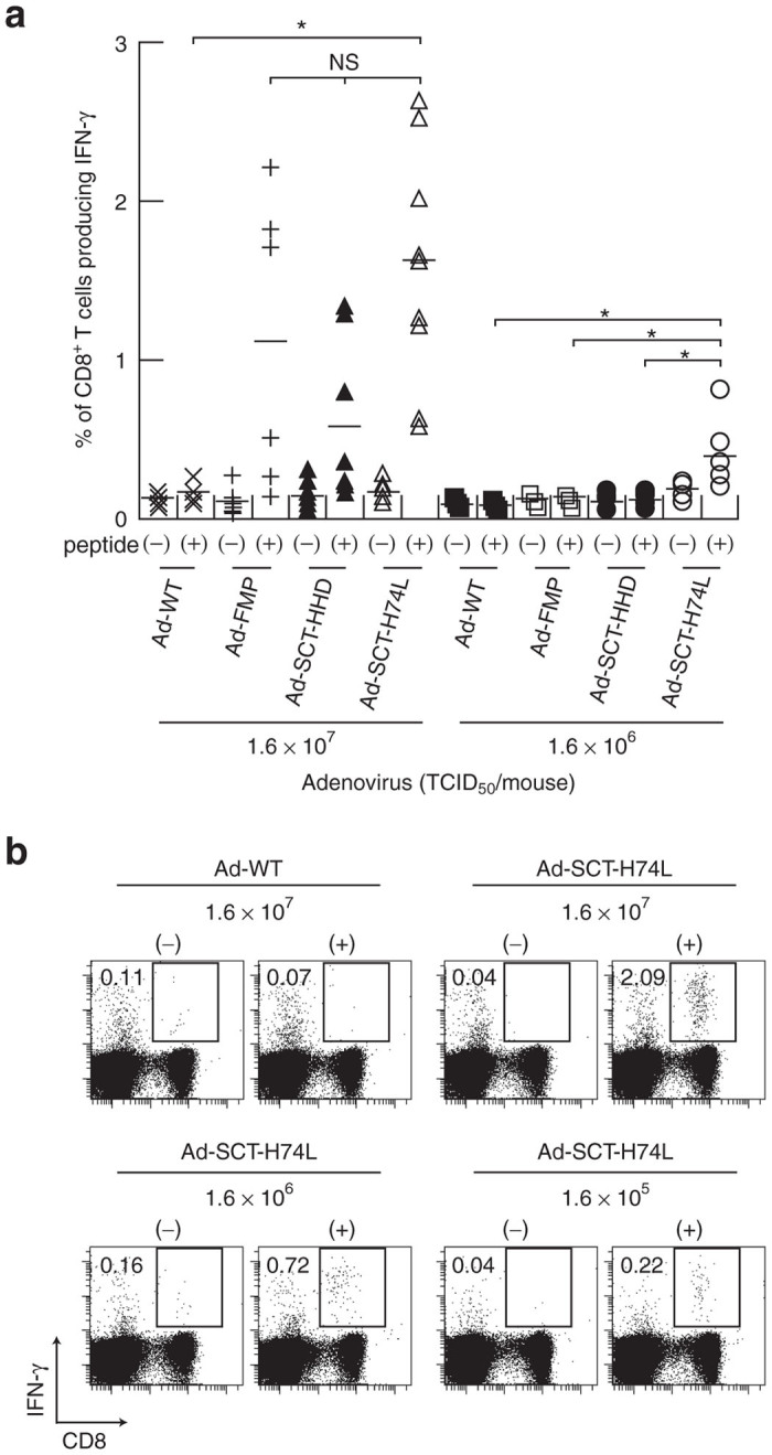
Intracellular IFN-γ staining of FMP-specific CD8+ T cells in mice immunized with Ad-SCT-H74L. HHD mice (five to nine per group) were immunized with Ad-WT, Ad-FMP, Ad-SCT-HHD or Ad-SCT-H74L at various inoculation doses. After 1 week, splenic lymphocytes were prepared, and stimulated with (+) or without (−) FMP58–66 for 5 hours. Cells were then stained for their surface expression of CD8 and their intracellular expression of IFN-γ. The data indicate the percentages of INF-γ-producing cells within CD8+ T cells. (a) Each symbol represents an individual mouse. Horizontal bars represent the mean. *P < 0.05; NS, not significant, One-way analysis of variance. (b) Data are representative in mice immunized with Ad-WT or Ad-SCT-H74L. The numbers shown indicate the percentages of INF-γ-secreting cells within CD8+ T cells.