Figure 3.
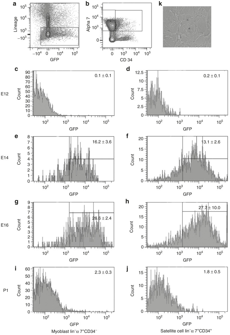
FACS analysis. Myf5nlacZ/+ mice underwent Intravascular injection with AAV 9 carrying GFP as a reporter gene at E12, E14, E16, and P1. Following myoblast prep of injected legs, cells were analyzed by FACS. Lineage negative cells were gated upon (a). These cells were analyzed for α 7 integrin (y axis) and CD34 (x axis) (b) and revealed two peaks: lineage negative, α 7 positive, and CD34 positive satellite cells and lineage negative, α 7 positive and CD34 negative myoblasts. Animals injected at E12, E14, E16, and P1 were analyzed for GFP positive satellite cells and myoblasts. (c,d) E12 injected myoblasts and satellite cells revealed 0.1 ± 0.1 and 0.2 ± 0.1% GFP transduction respectively. (e,f) E14 injected myoblasts and satellite cells revealed 16.2 ± 3.6 and 13.1 ± 2.6% GFP transduction respectively. (g,h) E16 injected myoblasts and satellite cells revealed 26.6 ± 2.4 and 27.7 ± 10.0% GFP transduction respectively. (i,j) P1 injected myoblasts and satellite cells revealed 2.3 ± 0.3 and 1.8 ± 0.5% GFP transduction respectively. (f) The GFP positive Satellite cells were injected into irradiated Rag mice and following engraftment, injury with notexin and healing failed to show GFP positive muscle fibers but (k) X-gal (blue) cells could be found in the satellite cell position.

