Figure 3.
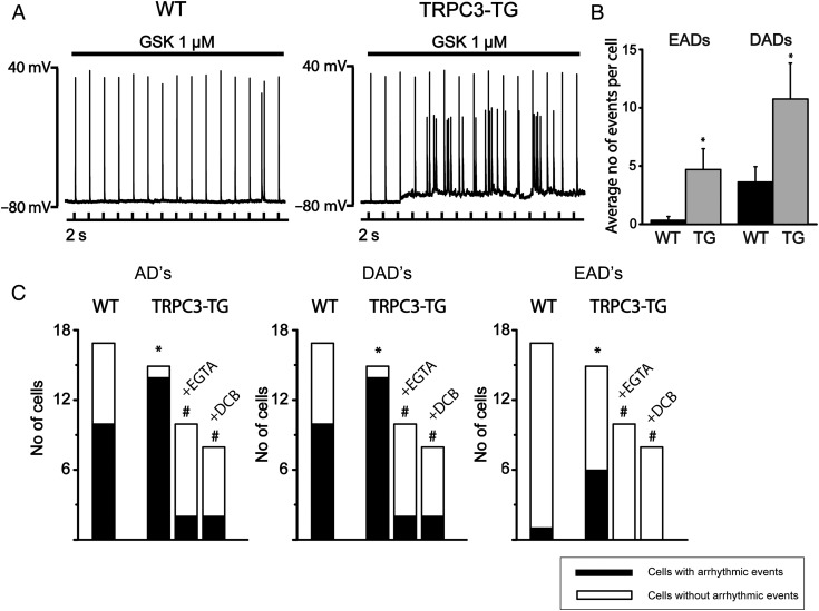
Arrhythmic activity in cardiomyocytes in response to TRPC3 activation requires rise of intracellular Ca2+ and NCX1 function. Action potentials were recorded from single myocytes upon intracellular current injection (1–2 nA amplitude, 2–4 ms duration) at a pacing cycle length of 2 s.28 (A) Continuous AP recordings displayed prominent arrhythmic afterdepolarizations (EADs and DADs) in TRPC3-TG myocytes during GSK infusion; WT (left panel), TRPC3-TG (right panel). (B) Frequency of arrhythmic events (number of EADs and DADs during 10 min application of GSK). Both EADs (4.7 ± 1.79 TG; n = 15; N = 3 vs. 0.3 ± 0.3 WT; n = 17; N = 4; P < 0.05) and DADs (10.75 ± 3.08 TG vs. 3.63 ± 1.32 WT; P < 0.05) were significantly increased in TRPC3-TG myocytes. Statistical significance analysed by Wilcoxon–Mann–Whitney test. (C) Incidence of afterdepolarizations (AD, left), delayed ADs (DAD, centre), and early ADs (EAD, right): WT compared with TRPC3-TG cells ± buffered to diastolic levels by 11 mM EGTA (n = 10; N = 3) or in the presence of the NCX1 inhibitor DCB (10 µM; n = 8; N = 3). Black bars represent population of cells displaying arrhythmic events during 10 min (n > 3). * indicates statistical significance (P < 0.05) in comparison to WT conditions, sharp (#) indicates (P < 0.05) in comparison to untreated TRPC3-TG myocytes. Statistical significance analysed by Fisher’s exact test.

