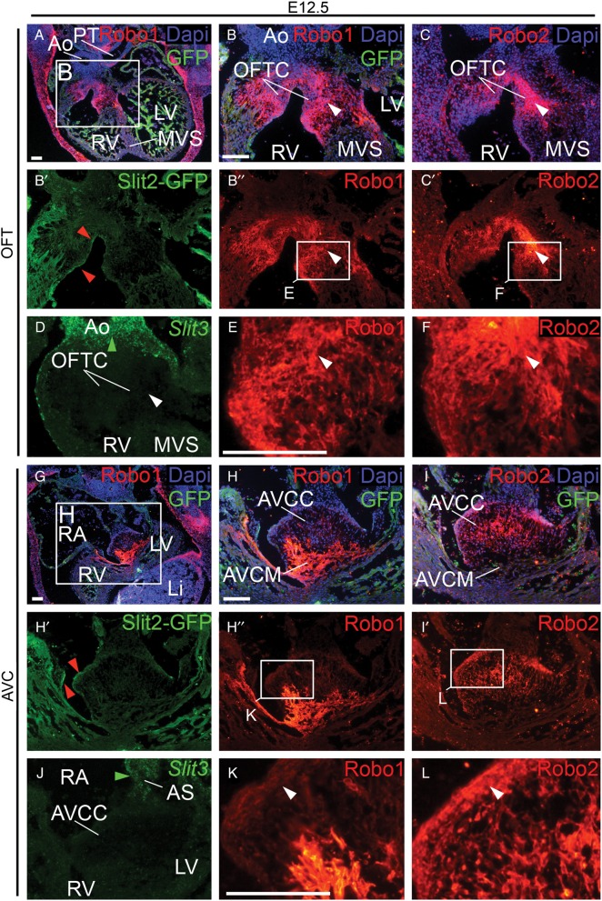
Figure 1.
Slit and Robo expression patterns in and surrounding the cardiac cushions. (A–L) immunohistochemistry (Robo1, Robo2, and Slit2-GFP), DAPI, and in situ hybridization (Slit3) staining at E12.5 in the outflow tract and atrioventricular cushion regions. Red arrowheads indicate the expression of Slit2 in the endocardium lining the cushions. (E, F, K, and L) show details of Robo1 and 2 expression in the indicated cushion regions. White arrowheads point to the region where both Robo1 and Robo2 are expressed. (D and J) Green arrowheads point to Slit3 expression in the outflow tract vessels and atrial septum, while Slit3 is not detectable in the cushions. Per stage for all genes analysed, n ≥ 3 embryos. Ao, Aorta; AVC, atrioventricular canal; AVCC, atrioventricular cushion; AVCM, atrioventricular canal myocardium; GFP, green fluorescent protein; Li, liver; OFT, outflow tract; OFTC, outflow tract cushion; MVS, membranous ventricular septum; PT, pulmonary trunk; RA, right atrium; R/LV, right/left ventricle. Scale bars depict 100 µm.

