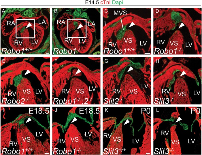
Figure 2.
Disrupted Slit-Robo signalling results in membranous ventricular septum defects. (A–L) immunohistochemistry for cardiac Troponin I (cTnI) and DAPI on Robo1+/+ (A, C, and I), Robo1−/− (B, D, and J), Robo2−/− (E), Robo1−/−;Robo2−/− (F), Slit2−/− (G), Slit3+/+ (K) and Slit3−/− (H and L) hearts. The valves and the membranous ventricular septum are visible as green DAPI staining. White arrowhead points to the presence or absence of the membranous ventricular septum (see Table 1 for numbers of embryos analysed). VS, (muscular) ventricular septum. For other abbreviations, see the legend of Figure 1. Scale bars depict 100 µm.

