
Figure 3.
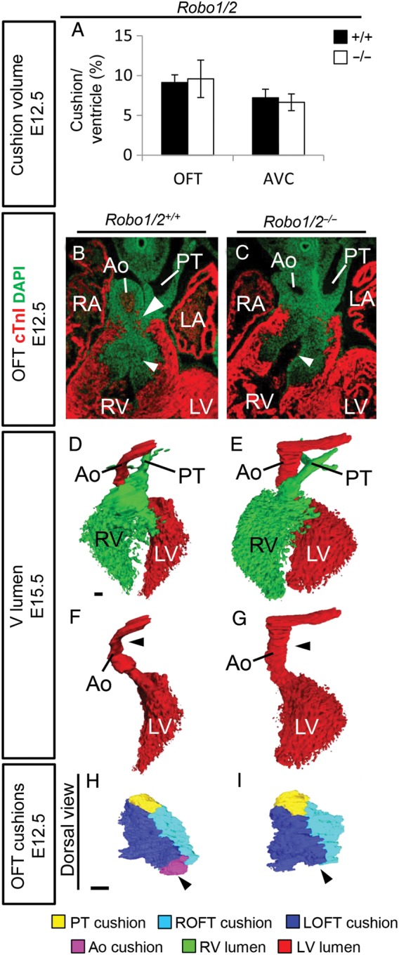
Defects in cushion formation in Robo1/2 double mutants. (A) Measurements of the OFT and AVC cushion volume corrected for ventricular volume (n = 3; OFT, P = 0.51; AVC, P = 0.27; Mann–Whitney U test). (B) Immunohistochemistry for cardiac Troponin I (cTnI) and DAPI on Robo1+/+;Robo2+/+, and Robo1−/−;Robo2−/− mutants. White arrowheads indicate increased space between OFT cushions in the mutant at E12.5. (D–G) Three-dimensional reconstructions of the ventricular lumen and outflow tract vessels of E15.5 Robo1+/+;Robo2+/+ and Robo1−/−;Robo2−/− hearts showing reduced rotation of the aorta in the mutant (black arrowheads), most clearly visible after removing the right ventricle and pulmonary trunk (F–G). (H–I) Three-dimensional reconstructions of the outflow tract cushions of E12.5 Robo1+/+;Robo2+/+ and Robo1−/−;Robo2−/− embryos showing the absence of the aortic posterior cushion in the Robo1/2 mutant. n = 3. For abbreviations, see the legend of Figure 1. Scale bars depict 100 µm.